Invasive Endoscopic Spine Surgery
INTERVENTION THROUGH A NATURAL CHANNEL The latest in science and technology in treating the problem of Lumbosciatica.
The intervertebral disc is often wrongly blamed and targeted as the cause of lumbar pain. It has now been proven that pain is often due to pathology that occurs in the epidural space.
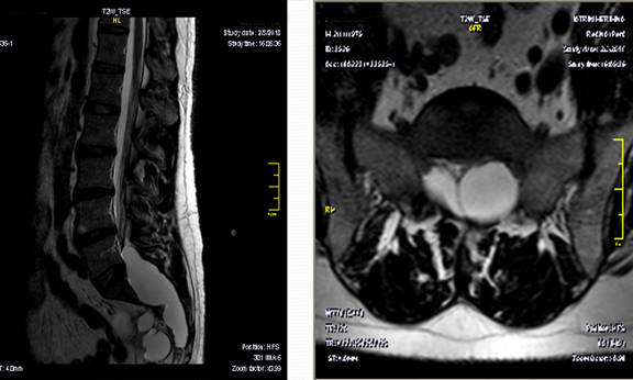
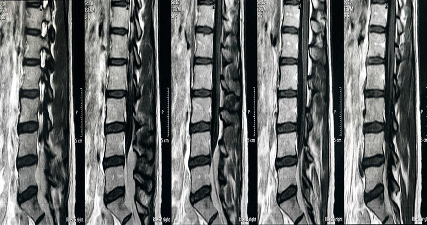
MRI as a diagnostic tool cannot distinguish inflammation and often gives insufficient (unreliable) and false data.
Classic spinal surgical techniques often fail or cause further problems due to the creation of scar tissue and destruction of healthy spinal structures.
All other invasive techniques refer to one level of the spine and are not diagnostic.
THE TECHNIQUE TREATS MOST CAUSES OF BACK PAIN
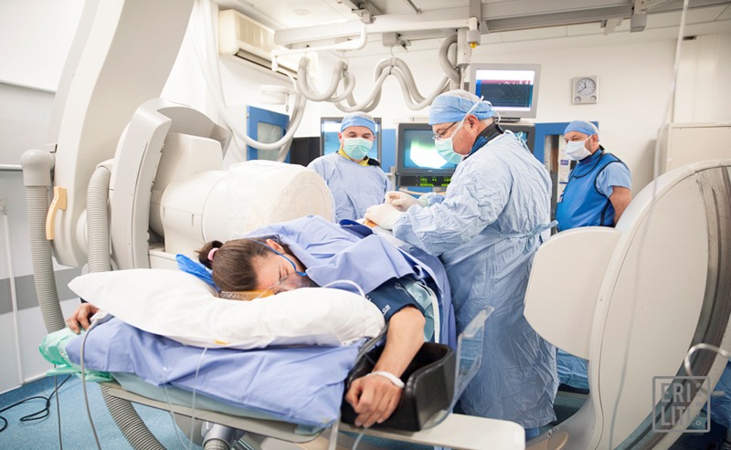
The procedure takes place in the interventional radiology room. It lasts 30-40 minutes and the patient leaves the hospital after a few hours. Most patients return to work the next day.
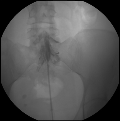
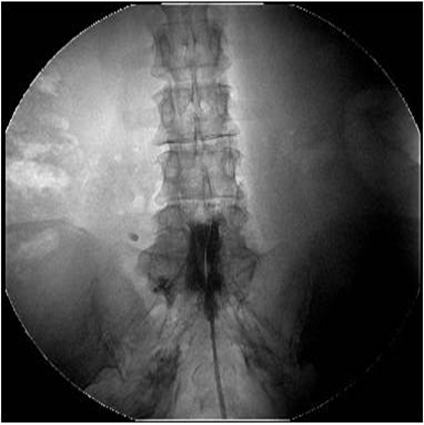
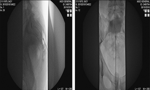
During the procedure, pathologies that cannot be detected by MRI are revealed and treated. The patient, under local anesthesia and light intoxication, communicates with the doctor throughout the procedure. The doctor inserts a thin navigable catheter with a camera through the sacral fissure into the spinal canal. Under the patient’s guidance, the symptomatology is reproduced. X-rays identify the height of the lesion and endoscopy the underlying pathology.
When the problem is identified, the appropriate treatment is applied (adhesions removal, nerve root release, ablation – shrinkage of structures using LASER)
ADVANTAGES OF MICROINVASIVE ENDOSCOPIC NEUROPLASTY – SPINE SURGERY
Invasive Endoscopic Neuroplasty of the OMSS
The diagnostic study and treatment, mainly of chronic degenerative diseases of the OMSS and failed surgery syndrome, is carried out in 3 stages:
1. Dynamic study
ENDOSCOPIC ACCESS TO THE SPINAL TUBE AND WOUNDS
DYNAMIC STUDY – ENDOSCOPE NAVIGATION
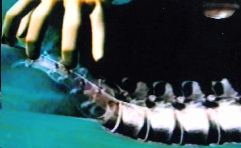
Mechanical irritation of the affected root reproduces the symptoms and has a positive predictive value ranging from 87%-100%.
MECHANICAL IRRITATION OF THE NERVE ROOT
2. Imaging study
SPINAL TUBE NESTIA
3. Endoscopic study
ENDOSCOPIC ANATOMY
(LUMBAR ROOT WITHIN THE SPINAL TUBE)
LUMBAR ROOT WITHIN THE WOUND
INFLAMED NERVE ROOT
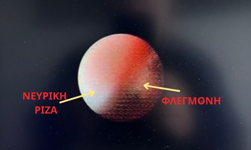
INFLAMED NERVE ROOT
VENOUS STATUS
DENTIFYING THE MENING SAC
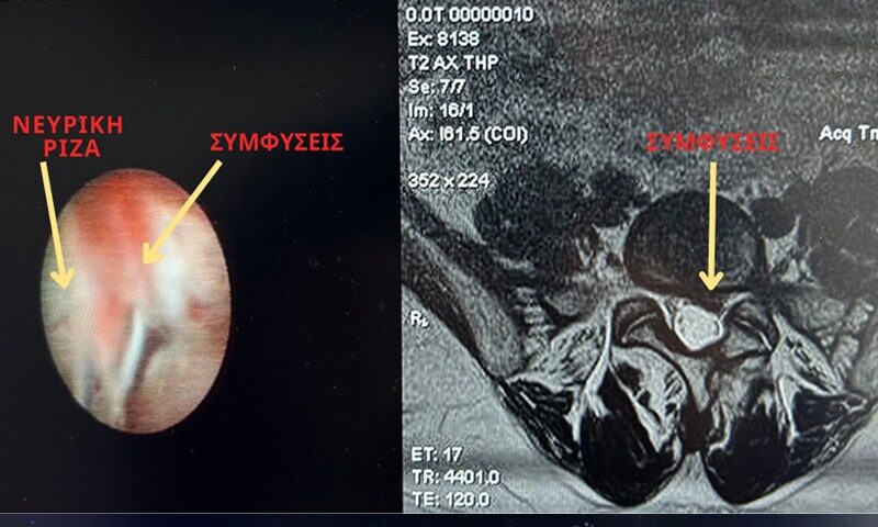
WOUND NARRATION
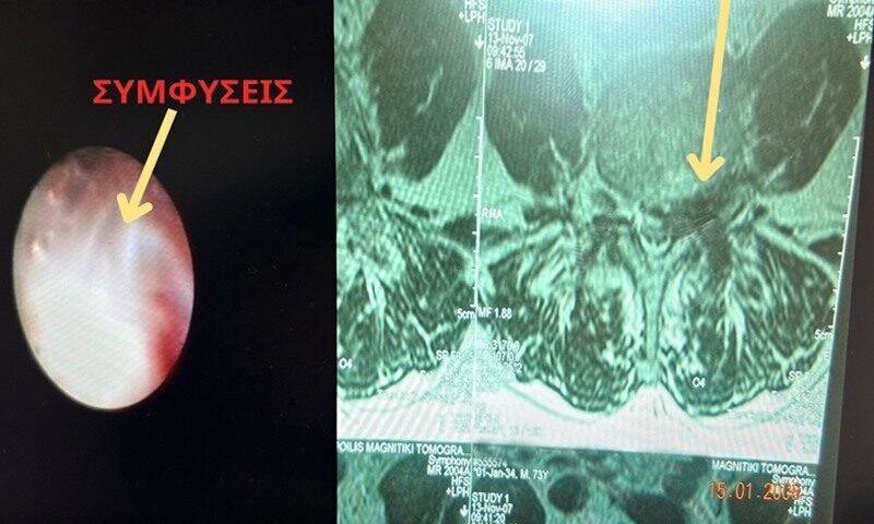
LYSIS OF ADHESIONS WITH HOLMIUM LASER
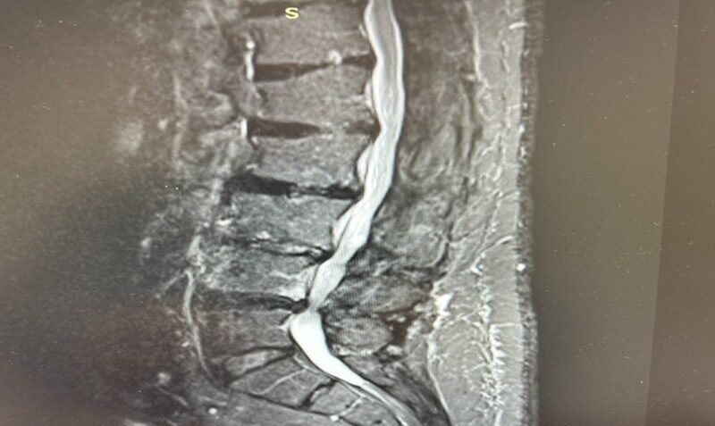
NEBULA PRESURGERY
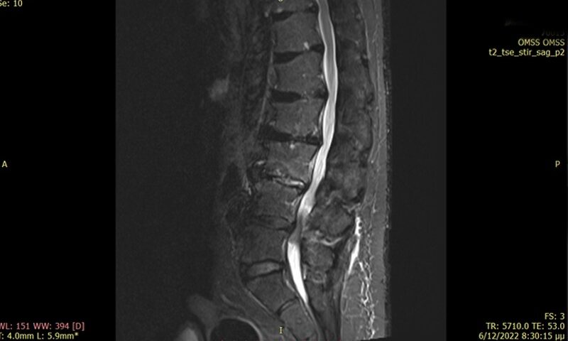
POSTOPERATIVE NEBULA
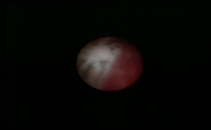
ARTERIOVENOUS DYSPLASIA OF THE MENINGA SCLES
TRANSTUNAL SCREW WITHIN A SPINAL TUBE
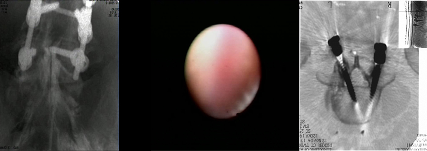
Interventional Endoscopic Surgery of the OMSS
FREE DISC MATERIAL
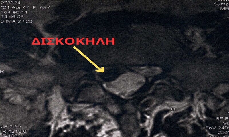
PERINEURAL CYSTS
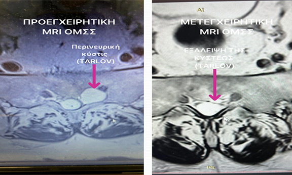
GIANT PERINEURAL CYSTS
DISCOHERAL O5-I1 – PERINEURAL CYSTS I3
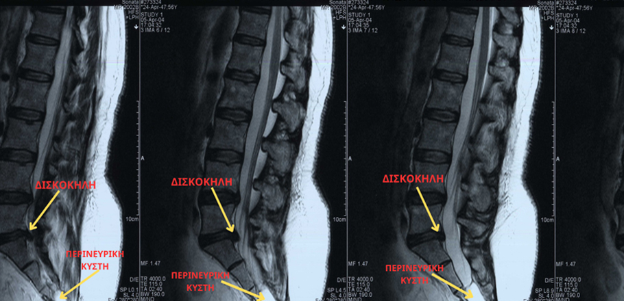
JOINT CAPSULE CYSTS – PRESURGERY
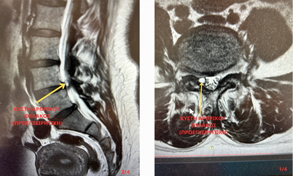
JOINT CAPSULE CYSTS – POST-SURGERY
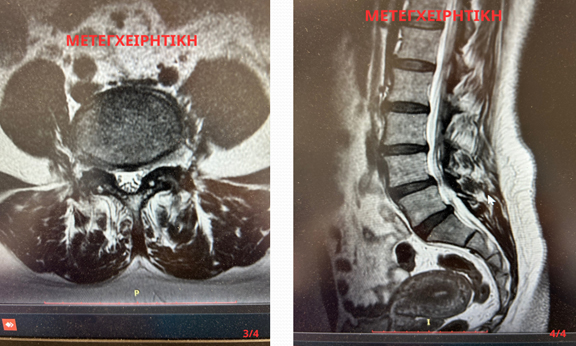
JOINT CAPSULE CYSTS
SPINAL TUBE NESTIA
FINAL FILAMENT
PSEUDOMENINGOCELE
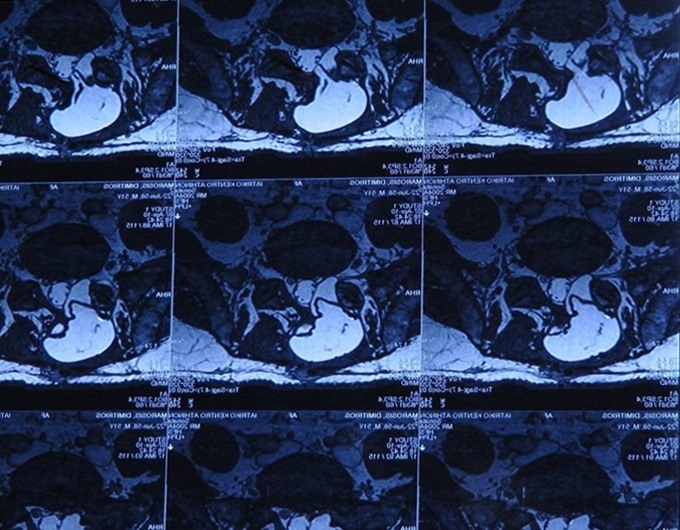
EPISCRECIDE LIPOMATOSIS
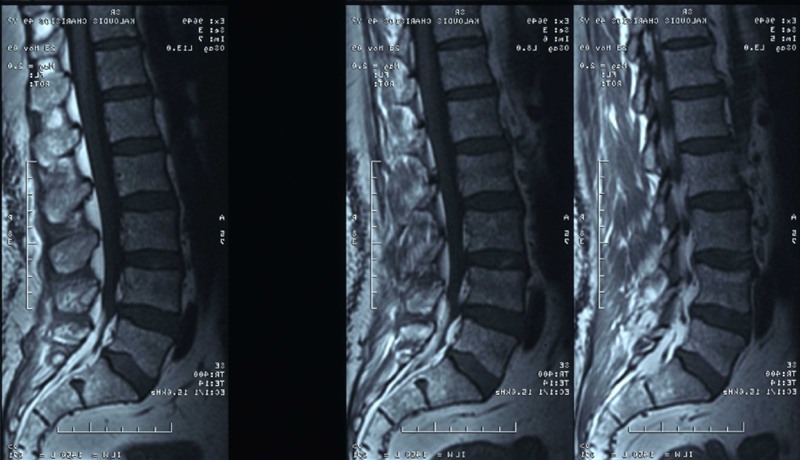
SPACE-INCESSIVE PROCESSING OF OMSS
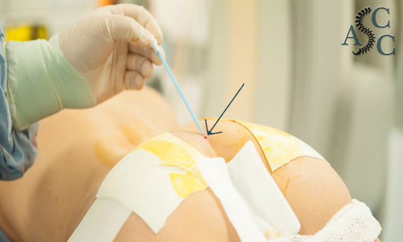
FINAL RESULT SKIN INCISION 1-2 mm
ENDOSCOPIC NEUROPLASTY 3D ANIMATION
The concept of Minimally Invasive Endoscopic Spine Surgery aims to achieve better clinical results compared to classical surgery because in this way:
The detailed evaluation of clinical, endoscopic and imaging data and the correct selection of patients is the true surgical art and not the simple application of a specific surgical technique.
At ADVANCED SPINAL CARE CLINIC, surgeries are performed on the entire Spine with the appropriate techniques (minimally invasive, endoscopic, classic) depending on the pathology.

